For manufacturers in electronics, aerospace, and advanced materials like metals, alloys, and composites, Scanning Acoustic Microscopy (SAM) offers a powerful quality control tool, ensuring structural integrity, reliability, and performance—all without damaging a single component. SAM uses high-frequency ultrasound to inspect and characterize internal features of materials, detecting cracks, voids, inclusions, and delaminations that could compromise performance.
SAM is a powerful non-invasive and non-destructive method for inspecting internal structures in optically opaque materials. Depth-specific information can be extracted and applied to create two- and three-dimensional images without the need for time-consuming tomographic scan procedures and more costly X-rays.
Today, SAM facilitates the detection of much smaller defects than previously possible.
“Advanced, phased array SAM systems make it possible to move to a higher level of failure analysis because of the level of detection and precision involved. In the past, detecting a 500-micron defect was the goal; now it is a 50-micron defect. With this type of testing, we can inspect materials and discover flaws that were previously undetected,” said Hari Polu, President of OKOS, a Virginia-based manufacturer of industrial SAM ultrasonic non-destructive testing systems. The company serves the electronics manufacturing, aerospace, and metal/alloy/composite manufacturers, and end-user markets.
For electronics manufacturers, SAM is indispensable for inspecting microchips, bonded wafers, and underfills, where failure is not an option. Aerospace firms rely on it to identify subsurface flaws in lightweight composites or high-performance alloys, ensuring safety in flight-critical applications. In metals and composites, it verifies adhesion quality and detects fatigue or internal corrosion early, saving money and lives.
However, to unlock the full power of SAM, manufacturers need the right system. Toward this goal, the most effective SAM systems are built on a triumvirate of high-performance components: transducers, digitizers, and software.
Optimizing SAM with Critical Components
Polu explains how transducers, digitizers, and software seamlessly work together in Scanning Acoustic Microscopy to benefit manufacturers.
“Transducers generate and receive the ultrasound signals, acting as the system’s ‘eyes.’ Digitizers convert high-frequency acoustic signals into precise digital data for analysis. Software brings it all together, enabling real-time visualization, defect identification, and actionable reporting. When these three elements work in harmony, SAM becomes more than a quality control step—it becomes a competitive advantage,” says Polu.
Transducers
A Scanning Acoustic Microscope operates by utilizing a transducer that converts electrical energy into highly focused, ultrahigh-frequency sound waves. These waves are directed to a precise point on the target object, enabling internal inspection with exceptional accuracy. The shape of the transducer’s lens and the frequency of the sound waves determine both the focal length and the resolution of the scan.
As the sound waves interact with the internal features of the material, they reflect back to the transducer, which then converts the returning acoustic signals into voltage. This returning analog signal is subsequently amplified by a pulser/receiver and digitized for further analysis. All ultrasonic scanning systems rely on this essential dual function—signal generation and detection via at least one transducer—to perform precise, non-destructive evaluations of internal structures.
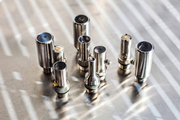
Transducers come in a variety of sizes and shapes for different applications. Some require direct contact with a material to operate; others use an air gap or are immersed in a fluid, usually water, in order to better transmit the sound wave through a material. OKOS offers a large variety of transducers up to 300 MHz for different applications and can custom engineer transducers for specific applications to suit specific needs.
According to Polu, the OEM offers four general types of transducers (Epoxy, PVDF, Delay Line, Phased Array), each of which has advantages for certain applications:
Epoxy tipped transducers tend to be less than 30 MHz and are useful for imaging thick samples or samples with very attenuative material. These often have the largest focal lengths.
PVDF transducers use a gold-tipped exposed element for high-frequency imaging, operating between 35 MHz and 75 MHz. These are ideal for thin, attenuative materials like silicon-based chips. Focal lengths typically range from 0.25 to 1.5 inches, enabling precise internal inspection.
Delay Line transducers are quartz lens tipped transducers with internal crystals manufactured to a precise thickness to control frequency. These transducers can range from 35 MHz to 300 MHz, have the best depth of field, and can have custom focal lengths.
Phased array transducers use multiple elements, unlike the single-element design of standard types, and can be curved to improve scanning over contoured surfaces. Multiple elements sweep the sample simultaneously, enabling faster scans. Constructive interference allows real-time focal length adjustment for optimal imaging. These transducers typically operate at 20 MHz or below.
Multiple transducers speed scanning
Unlike conventional Scanning Acoustic Microscopy systems that utilize a single-element transducer, phased array systems employ multiple transducers that can be combined to scan the sample simultaneously.
In a phased array system, multiple elements can be activated either simultaneously or sequentially to synthesize a focused acoustic beam. The number of transducer elements incorporated into the array varies significantly depending on the specific application and system design. Common configurations typically include arrays with 16, 32, 64, 128, or 256 elements.
“A conventional 5 MHz sensor could take up to 45 minutes to inspect an 8–10-inch square or disc alloy. Today, however, an advanced phased array with 64-128 sensors and innovative software to render the images can reduce inspection time to five minutes, with more granular detection of small impurities or defects,” says Polu.
A phased array scanning system consists of multiple ultrasound transducer elements arranged in an array. Each element within the array is independently controlled with respect to the timing (phase) and amplitude of excitation. This configuration allows for electronic steering and focusing of the ultrasound beam by adjusting the timing and amplitude applied to each element.
Phased array SAM systems offer significant advantages for applications that demand high-throughput inspection. These systems are particularly well-suited for non-destructive evaluation of composites, bonded structures, and electronic assemblies. They also support real-time imaging with adjustable depth of focus, which enhances their effectiveness in assessing internal features at various depths within the material.
“To produce an image, samples are scanned point by point and line by line,” explains Hari. “Scanning modes range from single layer views to tray scans and cross-sections. Multi-layer scans can include up to 50 independent layers. Depth-specific information can be extracted and applied to create two-and three-dimensional images without the need for time-consuming tomographic scan procedures or costly X-ray equipment. The images are then analyzed to detect and characterize flaws such as cracks, inclusions, and voids.”
According to Polu, SAM can also be custom designed to be fully integrated into high volume manufacturing systems. When high throughput is required for 100% inspection, ultra-fast single or dual gantry scanning systems are utilized along with 128 transducers for phased array scanning.
Digitizers
In a Scanning Acoustic Microscope, the digitizer takes the analog voltage signals received from the transducer—after amplification by the pulser/receiver—and converts them into digital format. This digital data is then used for image reconstruction and analysis, enabling accurate visualization of the internal features of the inspected object. The digitizer is critical for translating raw acoustic information into usable, high-resolution imaging.
Digitizers convert analog signals into digital form by sampling the input waveform at specific intervals, known as the sampling rate. A higher sampling rate captures more data points per second, allowing for a more accurate reconstruction of the original signal. To avoid distortion and preserve signal integrity, the sampling rate usually must be at least twice the highest frequency present in the signal, according to Polu.
“More data is generated as the sampling rate is increased, so the lowest sampling rate that can accurately reproduce the original signal will improve throughput,” says Polu.
Software
Software coordinates all the pieces of an ultrasonic scanning system like SAM. It interacts with the digitizer, motion control, and digital pulser/receivers in order to coordinate their operations. Software is used to adjust the position of the sample or the probe (transducer) in three-dimensional space, trigger the transducer, and process the resulting waveform data into 2D and 3D images.
As important as the physical and mechanical aspects of conducting a scan, the software is critical to improving the resolution and analyzing the information to produce detailed scans.
Multi-axis scan options enable A, B, and C-scans, contour following, off-line analysis, and virtual rescanning for a variety of materials. This results in highly accurate internal and external inspection for defects and thickness measurement via the inspection software.
Various software modes can be simple and user friendly, advanced for detailed analysis, or automated for production scanning. An off-line analysis mode is also available for virtual scanning.
Polu estimates that OKOS’ software-driven model enables them to drive down the costs of SAM testing while delivering the same quality of inspection results. Consequently, this type of equipment is well within reach of even modest testing labs.
The Bottom Line
In Scanning Acoustic Microscopy, performance depends not solely on the quality of individual components but on their effective integration.
Scanning Acoustic Microscopy achieves peak effectiveness when transducers, digitizers, and software operate in seamless coordination. Performance depends not only on the quality of individual components, but on how well they are integrated as a unified system.
When properly matched, these elements enable higher scanning speeds, enhanced defect detection at smaller scales, and improved imaging clarity. This level of performance allows manufacturers to identify issues early, enhance product quality, and maintain a competitive edge in industries where minor defects can result in significant consequences.
For more information, contact PVA TePla OKOS at fa@pvatepla.com or visit www.pvatepla-okos.com. OKOS is a wholly owned subsidiary of PVA TePla AG, Germany.

